Tomographic Imaging

Course given by
Prof. Dr.-Ing. Georg Schmitz
Course number
141223
Language
German
Credit Points
5
Hours per week
4
Click here to go to
Moodle
Contents
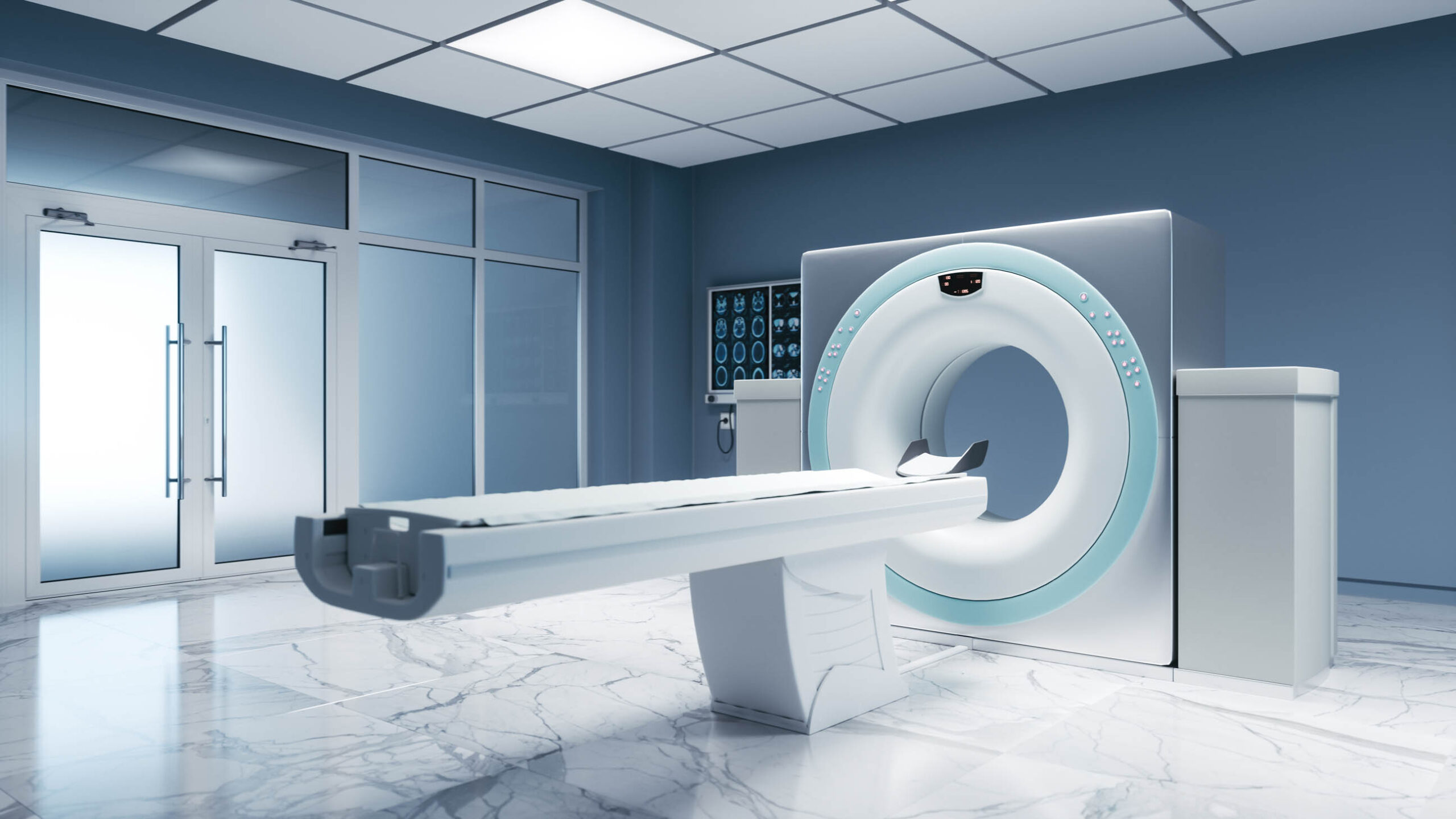
Using tomographic imaging techniques, image slices of tissue and bone structures can be reconstructed from projections, i.e. from measured, integral relationships of physical parameters. Computer tomography (CT) measures the penetration of X-rays through a volume to be imaged at different angles and reconstructs the X-ray attenuation coefficient. Magnetic resonance tomography (MR), on the other hand, uses nuclear magnetic resonance effects and images proton densities weighted by relaxation times. From the physical and mathematical basics to practically important reconstruction procedures, all steps from data acquisition to the image are taught.
Lectures
Room
ID 04/445
Lesson begins
08:15
Lesson ends
09:45
First lesson is on
Wednesday, 10.04.2024
Excercises
Room
ID 04/445
Excercise begins
10:15
Excercise ends
11:45
First excercise is on
Wednesday, 17.04.2024
Exam
Type of exam
Oral exam
Exam date
Individual Appointment
Duration of exam
30 minutes
Registration for the exam
FlexNow
Objectives
After successful completion of the module, students have knowledge of the most important tomographic diagnostic imaging procedures (X-ray computed tomography, magnetic resonance imaging). They know the basic technical components of the imaging systems under consideration and can explain how they work. They understand the basic physical effects (e.g. X-ray attenuation, nuclear magnetic resonance) and can discuss them. Students understand the theory of tomographic reconstruction (Fourier-Slice-Theorem, Fourier-Diffraction Theorem) and can derive and explain the structure and the achieved image quality of the different systems. They are able to implement known algorithms for image reconstruction and independently develop and evaluate new algorithms. Through the exercises in small groups, partly on computers, the students are able to apply what they have learned in a small team. They are able to explain their solutions and to present supporting arguments.
Requirements
none
Prior knowledge
Knowledge of system theory, Fourier transformation and signal processing, corresponding to those taught as basics in the lectures of the bachelor's program in Electrical Engineering and Information Technology.
Literature
- Oppelt (Hrsg.): Imaging Systems for Medical Diagnosis. 4. Auflage 2005. Publicis MCD Verlag. ISBN 3-89578-226-2.
- Buzug: Einführung in die Computertomographie. 1. Auflage 2004. Springer. ISBN 3-540-20808-9.
- Kak, Slaney: Principles of Computerized Tomographic Imaging. 1987, IEEE Press. ISBN 0-87942-198-3
- Vlaardingerbroek, den Boer: Magnetic Resonance Imaging. Springer, Berlin Heidelberg, 1996
- Liang, Lauterbur: Magnetic Resonance Imaging. A Signal Processing Perspective. 2000, IEEE Press. ISBN 0-7803-4723-4.
Miscellaneous
Registration is carried out via the E-Learning Portal Moodle of the Ruhr-Universität Bochum. The required information is provided in the first lecture.


