Superauflösende US Brustkrebs-Bildgebung

Weiterentwicklung der bewegungsmodellbasierten Ultraschall-Lokalisationsmikroskopie zur Unterstützung der Brustkrebsdiagnostik und Therapieüberwachung in Patienten
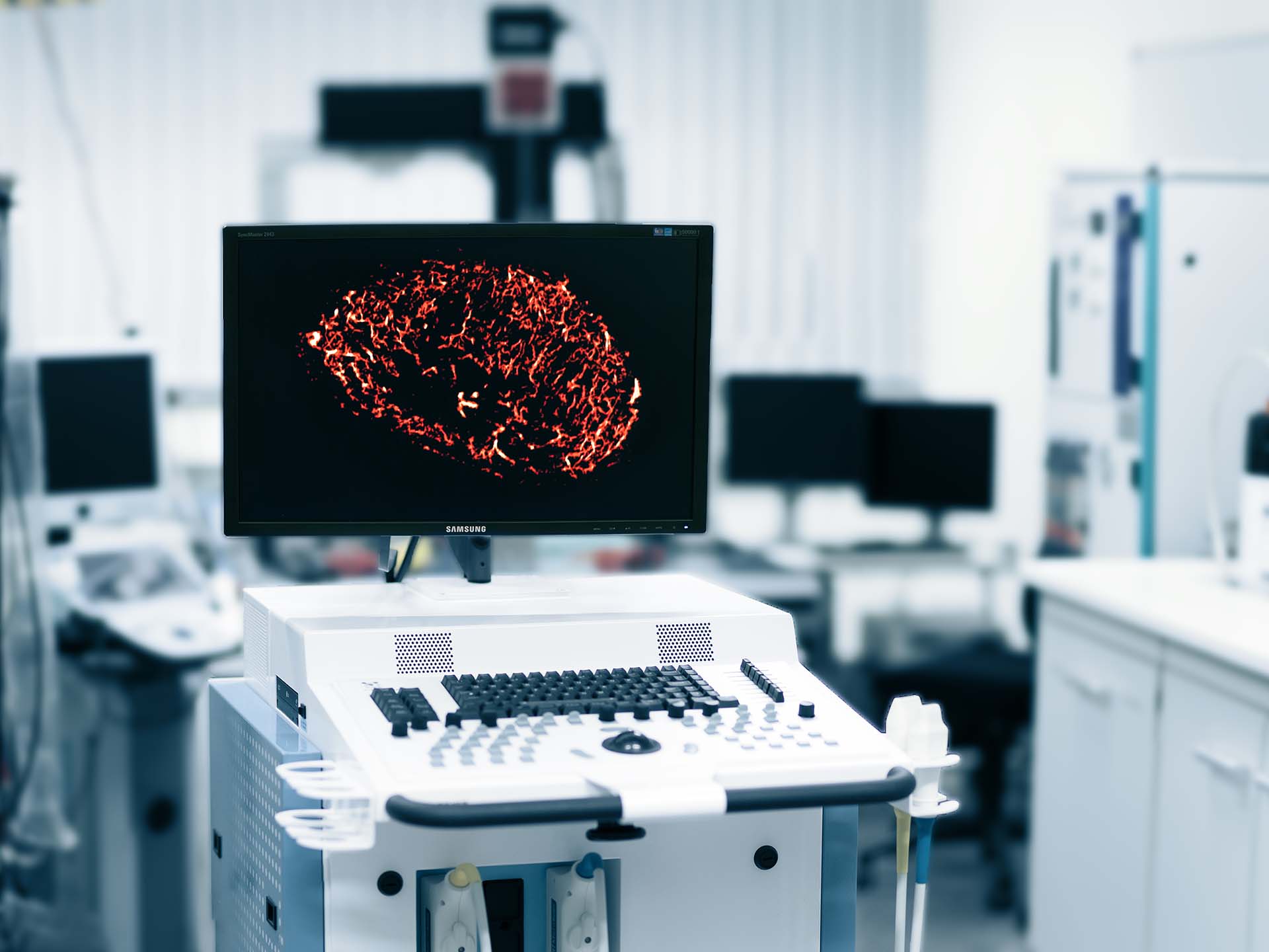
Ultraschallverfahren werden routinemäßig bei der Brustkrebsvorsorge und zur Beurteilung der Dignität verdächtiger Läsionen eingesetzt. Mit Unterstützung der DFG haben wir eine hochauflösende, kontrastverstärkte Ultraschallmethode entwickelt, die sogenannte Motion Model Ultrasonic Localization Microscopy (mULM), die einzelne Mikrobläschen in Videosequenzen verfolgt und Bilder des Gefäßsystems mit einer Auflösung jenseits der Beugungsgrenze erzeugt.
Die mULM-Methode ermöglicht eine genaue Beurteilung der Gefäßarchitektur in Tumoren, der einzelnen Blutgefäßgeschwindigkeiten und Flussrichtungen sowie die Quantifizierung des relativen Blutvolumens und der Perfusion. Zuvor haben wir an Mäuse-Xenograft-Tumoren gezeigt, dass diese Methode die Extraktion verschiedener morphometrischer Parameter ermöglicht und in Kombination mit einer radiomischen Analyse die automatische Unterscheidung verschiedener Tumortypen unterstützt.

In diesem Projekt wollen wir mULM und ultraschallbasierte Radiomik für die Anwendung am Patienten anpassen und die Technik zur 3D-Erfassung und -Verfolgung weiterentwickeln. In einer ersten Analyse klinischer Datensätze konnten wir bereits superauflösende Gefäßverläufe extrahieren. Für die robuste Anwendung im klinischen Umfeld ergaben sich jedoch einige technische Herausforderungen: Insbesondere die Bewegung des Patienten und die geringere Bildauflösung in Verbindung mit einer erhöhten Schichtdicke und nicht optimierten Injektionsprotokollen erlaubten nur die Verwendung kurzer Bildsequenzen und die Extraktion einer reduzierten Anzahl von Gefäßen.

In einem ersten Schritt wird daher die mULM-Methode verfeinert und für die Anwendung in der Brustbildgebung auf klinischen Ultraschallgeräten angepasst. Anschließend soll die 2D-mULM-Methode zur Überwachung des Ansprechens von Brustkrebs auf eine neoadjuvante Chemotherapie eingesetzt werden. Die Methode wird mit der konventionellen kontrastverstärkten Soographie verglichen. Darüber hinaus wollen wir die Methode von der 2D- zur vollständigen 3D-Erfassung mit einem volumetrischen Echtzeit-Scanner weiterentwickeln und beide Ansätze in einer kleinen Kohorte von Patienten mit Brusttumoren unterschiedlicher Dignität vergleichen. Um das volle Potenzial der mULM-Datenanalyse auszuschöpfen, werden Algorithmen entwickelt und implementiert, die eine automatische Extraktion und Clusteranalyse der vaskulären Merkmale ermöglichen. Zur automatischen Gruppierung der Patienten werden Ähnlichkeitsmessungen und Clusteranalysen durchgeführt. Somit wird dieses Projekt einen ersten Beitrag zur klinischen Umsetzung des multiparametrischen superauflösenden Ultraschalls leisten und den Weg für die ultraschallbasierte Radiomik-Analyse ebnen.

Projektinformationen

Projektbeginn
1. Juli 2013
Projektende
30. Juni 2023
Gefördert durch
German Research Foundation
Projektnummer
233312120

Veröffentlichungen

[1]
M. Lerendegui et al., “ULTRA-SR Challenge: Assessment of Ultrasound Localization and TRacking Algorithms for Super-Resolution Imaging,” IEEE Trans. Med. Imaging, vol. 43, no. 8, pp. 2970–2987, Apr. 2024, doi: 10.1109/TMI.2024.3388048.
[1]
C. Porte et al., “Ultrasound Localization Microscopy for Breast Cancer Imaging in Patients: Protocol Optimization and Comparison with Shear Wave Elastography,” Ultrasound in Medicine & Biology, vol. 50, no. 1, pp. 57–66, 2024, doi: 10.1016/j.ultrasmedbio.2023.09.001.
[1]
P. Hagemeyer, T. Lisson, S. Dencks, and G. Schmitz, “Fast Image Reconstruction in the Frequency Domain for Row-Column-Arrays,” presented at the IEEE International Ultrasonics Symposium (IUS), 2024, pp. 1–4.
[1]
T. Lisson, M. Fouad, J. Baier, A. Rix, F. Kiessling, and G. Schmitz, “A Comparative Study of Contrast Enhanced Ultrasound Imaging Using Deep Learning vs. Amplitude Modulation: An In-Vivo Investigation,” presented at the IEEE International Ultrasonics Symposium (IUS), 2024, pp. 1–4.
[1]
P. Hagemeyer, T. Lisson, S. Dencks, and G. Schmitz, “Fourier Diffraction Theorem for 3D Ultrasound Imaging with a Row-Column Array,” presented at the IEEE International Symposium on Biomedical Imaging (ISBI), 2024, pp. 1–5. doi: 10.1109/isbi56570.2024.10635345.
[1]
J. Sobolewski, S. Dencks, and G. Schmitz, “Influence of Image Discretization and Patch Size on Microbubble Localization Precision,” IEEE Trans. Ultrason. Ferroelectr. Freq. Control, 2024, doi: 10.1109/TUFFC.2024.3479710.
[1]
C. Porte et al., “Ultrasound Localization Microscopy for Cancer Imaging,” IEEE Transactions on Ultrasonics, Ferroelectrics, and Frequency Control, pp. 1–1, 2024, doi: 10.1109/tuffc.2024.3508266.
[1]
S. Dencks, T. Lisson, N. Oblisz, F. Kiessling, and G. Schmitz, “Ultrasound Localization Microscopy Precision of Clinical 3D Ultrasound Systems,” IEEE Transactions on Ultrasonics, Ferroelectrics, and Frequency Control, pp. 1–1, 2024, doi: 10.1109/TUFFC.2024.3467391.
[1]
T. Lisson, J. Salewski, S. Dencks, and G. Schmitz, “Resolution Improvement of ULM Images Applying a Rauch-Tung-Striebel Smoother,” in 2023 IEEE International Ultrasonics Symposium (IUS), Sep. 2023, pp. 1–4. doi: 10.1109/IUS51837.2023.10306605.
[1]
S. Dencks and G. Schmitz, “Ultrasound localization microscopy,” Zeitschrift für Medizinische Physik, Jun. 2023, doi: 10.1016/j.zemedi.2023.02.004.
[1]
S. Dencks, M. Piepenbrock, and G. Schmitz, “Velocity Filtering with a Median Filter Better Preserves Small Vessels for Ultrasound Localization Microscopy,” in 2021 IEEE International Ultrasonics Symposium (IUS), Sep. 2021, pp. 1–4. doi: 10.1109/IUS52206.2021.9593882.
[1]
M. Piepenbrock, D. Koretskaia, G. Schmitz, and S. Dencks, “3D Microbubble Localization with a Convolutional Neural Network for Super-Resolution Ultrasound Imaging,” in 2021 IEEE International Ultrasonics Symposium (IUS), Sep. 2021, pp. 1–4. doi: 10.1109/IUS52206.2021.9593473.
[1]
M. Piepenbrock, S. Dencks, and G. Schmitz, “Tissue Motion Estimation of Contrast Enhanced Ultrasound Images with A Stable Principal Component Pursuit,” in 2021 IEEE 18th International Symposium on Biomedical Imaging (ISBI), Apr. 2021, pp. 1642–1645. doi: 10.1109/ISBI48211.2021.9434155.
[1]
A. Rix et al., “Effects of contrast-enhanced ultrasound treatment on neoadjuvant chemotherapy in breast cancer,” Theranostics, vol. 11, no. 19, pp. 9557–9570, 2021, doi: 10.7150/thno.64767.
[1]
M. Piepenbrock, S. Dencks, and G. Schmitz, “Microbubble Tracking with a Nonlinear Motion Model,” presented at the 2020 IEEE International Ultrasonics Symposium (IUS), Sep. 2020, pp. 1–4. doi: 10.1109/IUS46767.2020.9251581.
[1]
S. Dencks, M. Piepenbrock, and G. Schmitz, “Assessing Vessel Reconstruction in Ultrasound Localization Microscopy by Maximum Likelihood Estimation of a Zero-Inflated Poisson Model,” IEEE Trans Ultrason Ferroelectr Freq Control, vol. 67, no. 8, pp. 1603–1612, Aug. 2020, doi: 10.1109/TUFFC.2020.2980063.
[1]
K. Christensen-Jeffries et al., “Super-resolution Ultrasound Imaging,” Ultrasound Med Biol, vol. 46, no. 4, pp. 865–891, Apr. 2020, doi: 10.1016/j.ultrasmedbio.2019.11.013.
[1]
M. Piepenbrock, A. Brieden, S. Dencks, and G. Schmitz, “Advancing the Feasible Microbubble Concentration in Super-Resolution,” presented at the IEEE International Ultrasonics Symposium (IUS), Oct. 2019, pp. 388–391. doi: 10.1109/ULTSYM.2019.8925761.
[1]
M. Piepenbrock, S. Dencks, and G. Schmitz, “Reliable Motion Estimation in Super-Resolution US by Reducing the Interference of Microbubble Movement,” presented at the IEEE International Ultrasonics Symposium (IUS), Oct. 2019, pp. 384–387. doi: 10.1109/ULTSYM.2019.8925566.
[1]
S. Dencks, M. Piepenbrock, and G. Schmitz, “Maximum-Likelihood Estimation to Assess the Degree of Reconstruction of Microvasculature from Super-Resolution US Imaging,” in 2019 IEEE International Ultrasonics Symposium (IUS), Oct. 2019, pp. 376–379. doi: 10.1109/ULTSYM.2019.8925640.
[1]
S. Dencks et al., “Clinical Pilot Application of Super-Resolution US Imaging in Breast Cancer,” IEEE Trans. Ultrason. Ferroelectr. Freq. Control, vol. 66, no. 3, pp. 517–526, 2019, doi: 10.1109/TUFFC.2018.2872067.
[1]
M. Piepenbrock, S. Dencks, and G. Schmitz, “Performance of Foreground-Background Separation Algorithms for the Detection of Microbubbles in Super-Resolution Imaging,” presented at the 2018 IEEE International Ultrasonics Symposium (IUS), Oct. 2018, pp. 1–4. doi: 10.1109/ULTSYM.2018.8579815.
[1]
S. Dencks et al., “Relative Blood Volume Estimation from Clinical Super-Resolution US Imaging in Breast Cancer,” presented at the 2018 IEEE International Ultrasonics Symposium (IUS), Oct. 2018, pp. 1–4. doi: 10.1109/ULTSYM.2018.8580013.
[1]
A. Rix et al., “Advanced Ultrasound Technologies for Diagnosis and Therapy,” J Nucl Med, vol. 59, no. 5, pp. 740–746, May 2018, doi: 10.2967/jnumed.117.200030.
[1]
T. Opacic et al., “Motion model ultrasound localization microscopy for preclinical and clinical multiparametric tumor characterization,” Nature Communications, vol. 9, no. 1, p. 1527, Apr. 2018, doi: 10.1038/s41467-018-03973-8.
[1]
B. Theek, T. Opacic, D. Mockel, G. Schmitz, T. Lammers, and F. Kiessling, “Automated Generation of Reliable Blood Velocity Parameter Maps from Contrast-Enhanced Ultrasound Data,” Contrast media & molecular imaging, vol. 2017, 2017, doi: 10.1155/2017/2098324.
[1]
S. Dencks, D. Ackermann, and G. Schmitz, “Evaluation of bubble tracking algorithms for super-resolution imaging of microvessels,” presented at the IEEE International Ultrasonics Symposium, Sep. 18, 2016.
[1]
I. Spivak et al., “Low-Dose Molecular Ultrasound Imaging with E-Selectin-Targeted PBCA Microbubbles,” Mol. Imaging Biol., vol. 18, no. 2, pp. 180–190, Apr. 2016, doi: 10.1007/s11307-015-0894-9.
[1]
D. Ackermann and G. Schmitz, “Detection and Tracking of Multiple Microbubbles in Ultrasound B-mode Images,” IEEE Trans. Ultrason. Ferroelectr. Freq. Control, vol. 63, no. 1, pp. 72–82, 2016, doi: 10.1109/TUFFC.2015.2500266.
[1]
D. Ackermann and G. Schmitz, “Super-resolution velocity estimation in microvessels using Multiple Hypothesis Tracking,” presented at the IEEE International Ultrasonics Symposium (IUS), Oct. 2015, pp. 1–4. doi: 10.1109/ULTSYM.2015.0219.
[1]
D. Ackermann and G. Schmitz, “Reconstruction of flow velocity inside vessels by tracking single microbubbles with an MCMC data association algorithm,” presented at the IEEE International Ultrasonics Symposium (IUS), Jul. 2013, pp. 627–630. doi: 10.1109/ULTSYM.2013.0162.
[1]
M. Siepmann et al., “Phase shift variance imaging - a new technique for destructive microbubble imaging,” IEEE Trans. Ultrason., Ferroelectr., Freq. Control, vol. 60, no. 5, pp. 909–923, 2013, doi: 10.1109/TUFFC.2013.2648.

Kontakt